PLL-g-PEG Adsorbed on PDMS

An Aqueous-Based Surface Modification of Poly(dimethylsiloxane) with Poly(ethylene glycol) to Prevent Biofouling

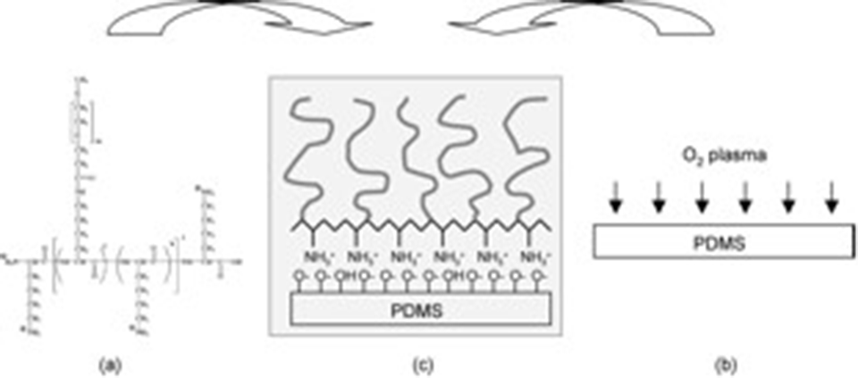
The application of poly(dimethylsiloxane) (PDMS) in biotechnology has been rapidly increasing, e.g., in microfluidic systems and microcontact print.  Despite many advantages, the surface properties of PDMS often demand further modification for successful application in biotechnology; nonspecific protein adsorption onto a PDMS surface is one of the most frequently encountered problems, e.g., in microfluidic systems.  We have employed a polycation-PEG graft copolymer, poly(L-lysine)-graft-poly(ethylene glycol) (PLL-g-PEG), which directly adsorbs onto the oxidized PDMS surface from aqueous solution. As shown in Figure 1, the polycationic nature of the PLL backbone leads to its electrostatic attraction onto a negatively charged oxidized PDMS surface in an aqueous environment at neutral pH, and thus, a densely packed PEG/water interface can be generated. We can thus avoid organic-solvent-based PEG-ylation approaches, which are known to swell the PDMS network structure.

Enlarged view: Figure 1. (a) The molecular structure of PLL-g-PEG. (b) Oxygen-plasma treatment of a PDMS surface. (c) Generation of PLL-g-PEG adlayer on top of oxidized PDMS surface driven by the electrostatic interaction between PLL backbone and negatively charged surface.
Figure 1. (a) The molecular structure of PLL-g-PEG. (b) Oxygen-plasma treatment of a PDMS surface. (c) Generation of PLL-g-PEG adlayer on top of oxidized PDMS surface driven by the electrostatic interaction between PLL backbone and negatively charged surface.

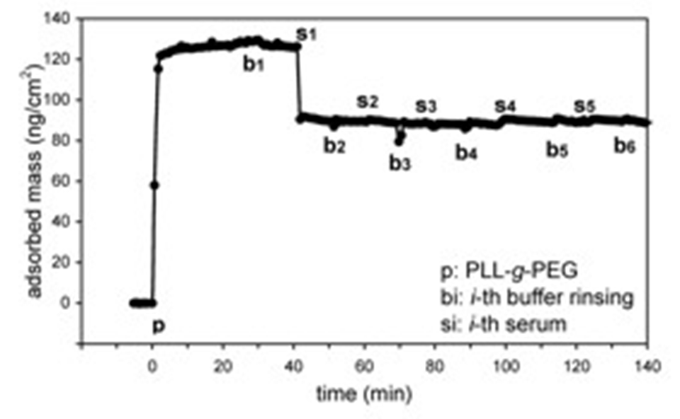
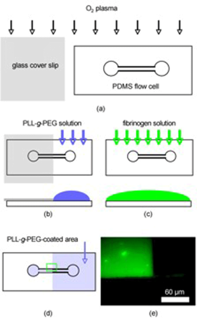
The PEG/aqueous interface generated in this way revealed a near-perfect resistance to nonspecific protein adsorption as monitored by means of optical waveguide lightmode spectroscopy (OWLS) (Figure 2) and Fluorescence Microscopy (Figure 3).

Enlarged view: Figure 2. Representative plot of the mass vs. time for the adsorption of PLL-g-PEG onto an oxygen-plasma-treated PDMS surface measured by OWLS (p; injection of PLL-g-PEG, bi; ith rinsing with HEPES buffer solution (i)1-11), si: ith injection of serum (i ) 1-10), only s1 to s5 and b1 to b6 are shown).
Figure 2. Representative plot of the mass vs. time for the adsorption of PLL-g-PEG onto an oxygen-plasma-treated PDMS surface measured by OWLS (p; injection of PLL-g-PEG, bi; ith rinsing with HEPES buffer solution (i)1-11), si: ith injection of serum (i ) 1-10), only s1 to s5 and b1 to b6 are shown).
Figure 3. Procedure for the fluorescence microscopy study employing a PDMS flow cell (a) a PDMS flow cell and a microscope glass cover slip were oxygen-plasma treated for 1 min, (b) the glass cover slip was attached to the PDMS surface
JavaScript has been disabled in your browser